File:SEM blood cells.jpg
Jump to navigation
Jump to search


Size of this preview: 482 × 600 pixels. Other resolutions: 193 × 240 pixels | 386 × 480 pixels | 617 × 768 pixels | 823 × 1,024 pixels | 1,800 × 2,239 pixels.
Original file (1,800 × 2,239 pixels, file size: 1.33 MB, MIME type: image/jpeg)
Captions
electron microscope image of blood
Captions
Summary[edit]
| Description |
English: This is a scanning electron microscope image from normal circulating human blood. One can see red blood cells, several white blood cells including lymphocytes, a monocyte, a neutrophil, and many small disc-shaped platelets. Red cells are nonnucleated and contain hemoglobin, an important protein that contains iron and allows the cell to carry oxygen to other parts of the body. They also carry carbon dioxide away from peripheral tissue to the lungs where it can be exhaled. The infection-fighting white blood cells are classified in two main groups: granular and agranular. All blood cells are formed in the bone marrow. There are two types of agranulocytes: lymphocytes, which fight disease by producing antibodies and thus destroying foreign material, and monocytes. Platelets are tiny cells formed in bone marrow and are necessary for blood clotting. Type: Black & White Print Русский : Это изображение нормально циркулирующей крови человека получено с помощью сканирующего электронного микроскопа. Можно видеть красные кровяные тельца, несколько белых клеток крови (в их числе лимфоциты, моноциты, нейтрофилы) и множество мелких дискообразных пластинок. Красные кровяные тельца содержат гемоглобин — важный белок, который содержит железо и позволяет клетке переносить кислород к другим частям тела. Также они переносят углекислый газ от периферических тканей в лёгкие, где тот после газообмена может быть выдохнут. Лейкоциты борются с инфекциями и на две основные группы: гранулярные и агранулярные. Все клетки крови образуются в костном мозге. Есть два типа агранулоцитов: лимфоциты, которые борются с болезнью, производя антитела и тем самым разрушая чужеродный материал, и моноциты. Тромбоциты представляют собой крошечные клетки, образующиеся в костном мозге, и необходимы для свертывания крови. Тип фото: чёрно-белая печать. |
||||||
| Date | Date Created: February 1982 | ||||||
| Source | Image and description: National Cancer Institute | ||||||
| Author | Bruce Wetzel (photographer). Harry Schaefer (photographer) | ||||||
| Permission (Reusing this file) |
|
||||||
| Other versions |
Derivative works of this file: |
||||||
.svg/64px-Great_Seal_of_the_United_States_(obverse).svg.png)
| Annotations | This image is annotated: View the annotations at Commons |
File history
Click on a date/time to view the file as it appeared at that time.
| Date/Time | Thumbnail | Dimensions | User | Comment | |
|---|---|---|---|---|---|
| current | 18:17, 3 February 2021 | | 1,800 × 2,239 (1.33 MB) | Tm (talk | contribs) | Reverted to version as of 20:27, 7 October 2006 (UTC) |
| 04:50, 10 November 2020 | 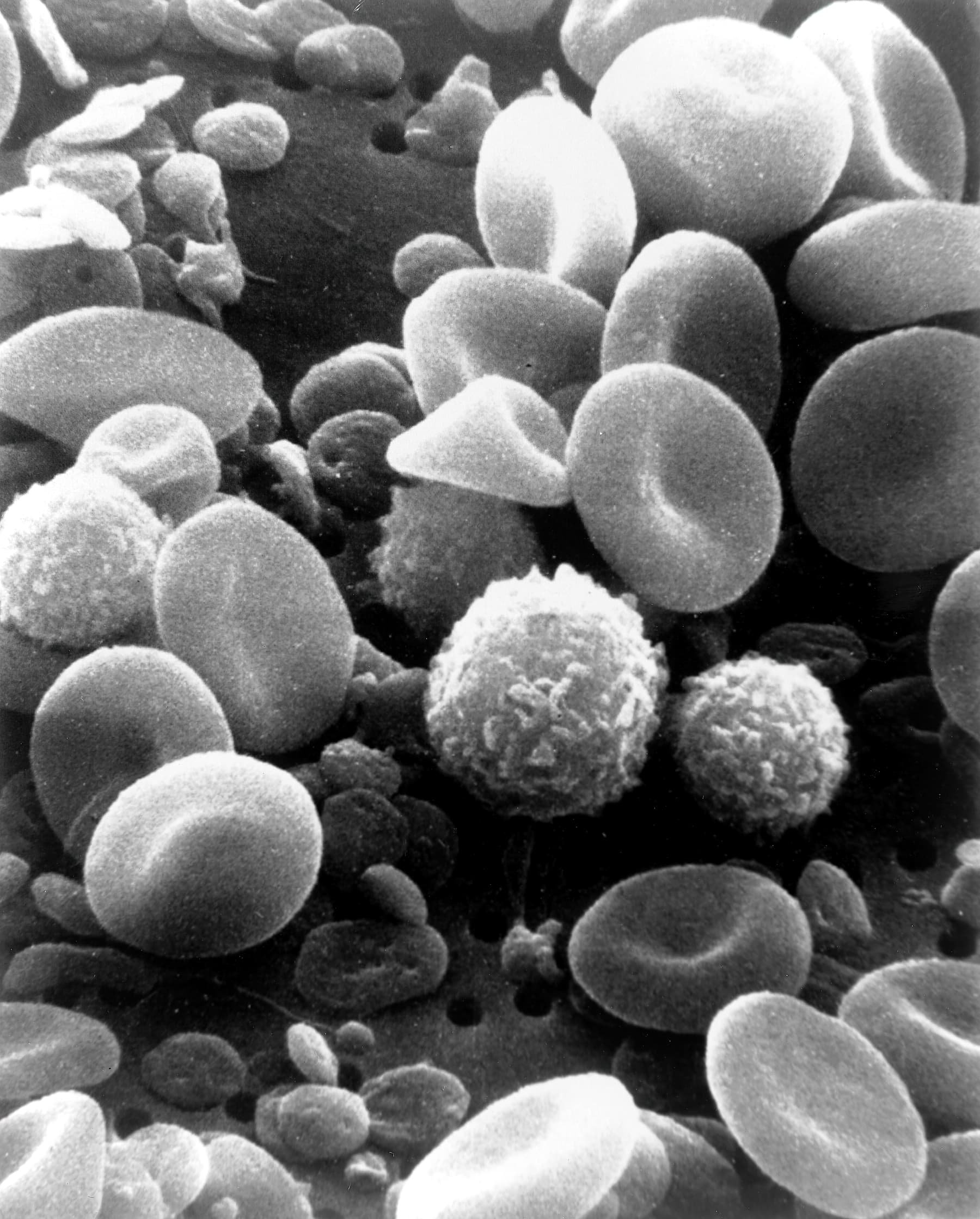 | 1,800 × 2,239 (309 KB) | Ratmanz (talk | contribs) | Optimized. | |
| 20:27, 7 October 2006 |  | 1,800 × 2,239 (1.33 MB) | DO11.10 (talk | contribs) | ||
| 03:00, 4 October 2006 |  | 1,800 × 2,239 (989 KB) | DO11.10 (talk | contribs) | {{Information |Description=This is a scanning electron microscope image from normal circulating human blood. One can see red blood cells, several white blood cells including lymphocytes, a monocyte, a neutrophil, and many small disc-shaped platelets. Red | |
| 01:09, 4 October 2006 | 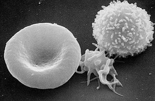 | 500 × 326 (36 KB) | DO11.10 (talk | contribs) | {{Information |Description= A three-dimensional ultrastructural image analysis of a T-lymphocyte (right), a platelet (center) and a red blood cell (left), using a Hitachi S-570 scanning electron microscope (SEM) equipped with a GW Backscatter Detector. |
You cannot overwrite this file.
File usage on Commons
The following 2 pages use this file:
File usage on other wikis
The following other wikis use this file:
- Usage on ar.wikipedia.org
- Usage on ar.wikiversity.org
- Usage on ast.wikipedia.org
- Usage on as.wikipedia.org
- Usage on az.wikipedia.org
- Usage on ba.wikipedia.org
- Usage on be-tarask.wikipedia.org
- Usage on be.wikipedia.org
- Usage on bg.wikipedia.org
- Usage on bn.wikipedia.org
- Usage on bn.wikibooks.org
- Usage on bs.wikipedia.org
- Usage on ca.wikipedia.org
- Usage on cs.wikipedia.org
- Usage on cv.wikipedia.org
- Usage on cy.wikipedia.org
- Usage on de.wikipedia.org
- Usage on dv.wikipedia.org
- Usage on el.wikipedia.org
- Usage on el.wiktionary.org
- Usage on en.wikipedia.org
- Immune system
- Scanning electron microscope
- Immunosuppression
- Orders of magnitude (volume)
- Lymphocyte
- Monocyte
- Talk:Scanning electron microscope
- Innate immune system
- User:Bishonen/Archive 11
- Wikipedia:Featured picture candidates/October-2008
- Wikipedia:Featured picture candidates/Human Blood Cells
- CFU-GEMM
- White blood cell
- User talk:Narayanese/Archive3
- Wikipedia:GLAM/NHMandSM/booklet
View more global usage of this file.